Speciality Contact Lenses 101
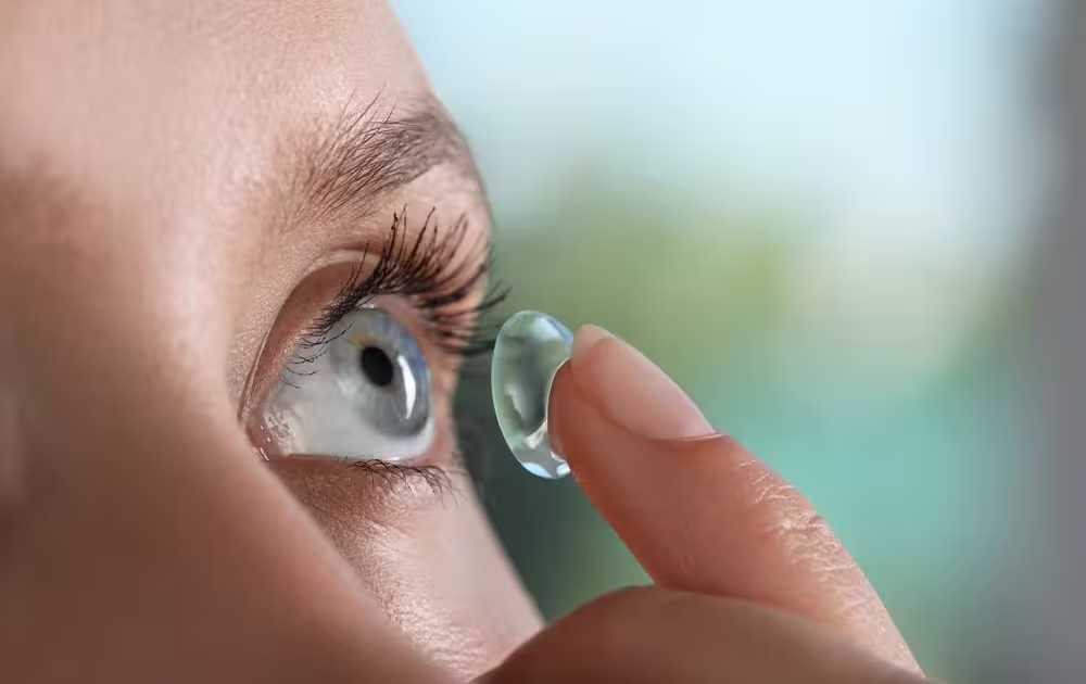
Contact Lens Most people have heard of soft disposable contact lenses. They either know someone who wears them or they wear these lenses themselves. There are different types of disposable lenses for frequent replacement (such as every day) or less frequent (every 3 months). They can correct astigmatism, presbyopia (the need for reading glasses) or even change your eye color. And even for high prescriptions, soft disposable lenses often fit the bill. For exceptionally high prescriptions, made-to-order soft contact lenses can be prescribed as well. But what about when soft contact lenses do not work? This is when specialty contact lenses may be the right choice. There are several types of specialty contact lenses, but today’s blog will focus on three main types: corneal rigid gas permeable (RGP), hybrid (soft and rigid in one lens), and scleral RGP. Corneal RGP lenses Corneal RGP lenses are small diameter lenses made of a rigid material that is permeable to oxygen. Unlike hard lenses of many decades ago, which did not let much oxygen into the eye, modern corneal RGP lenses have an array of oxygen permeable materials depending on the patient’s specific condition and oxygen needs. These lenses are placed directly on the cornea, and they are small in diameter such that no part of the lens edge will touch the sclera (white part of the eye). This type of lens can provide clearer vision than soft lenses for people with high prescriptions. It can also correct visual issues from conditions such as keratoconus, a condition which causes a warp in the cornea, corneal scarring, and other conditions which affect the shape of the cornea. The reason corneal RGP lenses can improve the vision in these conditions is because the rigid material masks any irregularities of the cornea under the lens. The lens actually floats on the eye, with tears filling in the void under the lens. This allows for a smooth anterior surface of the eye, which is ideal for refracting light to provide a clear image. A downside to this type of lens is the comfort. Because they have a smaller diameter than the cornea, every time a patient blinks, the lid will go over the edge of the lens. This can take several weeks for the patient to adjust to. Also, for more advanced cases of corneal irregularities, this type of lens may decenter and may come off of the eye. Hybrid lenses Similar to corneal RGP lenses, a hybrid lens has a rigid center; however it has a soft “skirt” surrounding the rigid center. This type of lens is used in the same conditions as corneal RGP lenses, but allows for better centration when a corneal RGP lens fails and is more comfortable at the outset. For some conditions, especially moderate to advanced keratoconus, a corneal lens may not center correctly on the eye, and it may even pop off the eye on occasion. In those scenarios, a hybrid lens will work well. The diameter of this lens can be quite a bit larger than a corneal lens, which allows for a better-fitting lens in conditions that affect the peripheral cornea. Depending on the type of hybrid lens prescribed, some brands need to be applied in a face-down position with preservative free saline in the bowl of the lens. Other types of hybrid lenses do not require saline for application. A downside to this type of lens are that the soft skirt can sometimes rip. Scleral lens Another type of specialty contact lens is a scleral lens. This is a large diameter RGP contact lens with no soft skirt. It is called a scleral lens because the edges of the lens rest mostly or entirely on the sclera. The advantages of this lens are the same as for the hybrid lens, but these lenses are more customizable, allowing for visual correction of a larger number of conditions. Additionally, they are typically very comfortable and stable on the eye. There is a steeper learning curve to application and removal of this type of lens (the lens is filled with preservative free saline and applied in a face-down position with a plunger; it is also removed with a plunger) but most patients have no problem learning how to handle these lenses with proper training. Scleral lenses can require more eye doctor visits than other lenses to optimize the lens fit and vision, but the payoff is definitely worth it, especially for patients who have severe corneal irregularities. The doctors in our offices who provide specialty contact lens care are Dr. Emmylou Swartz and Dr. Qian Yang. Book an Appointment
Understanding Cataracts
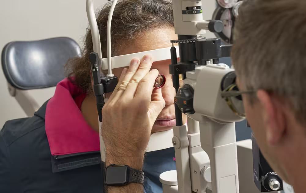
Cataracts are one of the most common causes of vision loss worldwide – but the good news is they are highly treatable. At Ophthalmology Associates, S.C., we specialize in helping patients see clearly again through advanced cataract evaluation and surgery. What Is a Cataract? A cataract occurs when the clear lens inside your eye becomes cloudy, making it difficult for light to pass through and focus properly on the retina. This can cause blurry vision, glare, faded colors, and difficulty seeing at night. Everyone eventually develops cataracts, and ophthalmologists usually start seeing some clouding of the lens in your eye around the age of 50-60. Cataracts typically develop slowly over time, and while they are most often related to aging, they can also occur earlier due to other factors. Who is at Risk for Cataracts? While everyone eventually develops cataracts, certain factors increase your risk and make them progress faster: Diabetes: High blood sugar can accelerate lens clouding. UV Exposure: Prolonged sunlight exposure without proper eye protection can lead to cataract formation. Smoking: Tobacco use increases oxidative stress in the eye, which can lead to cataracts. Certain Medications: Long-term steroid use, for example, may increase risk. Eye Injury or Surgery: Trauma or previous eye operations can lead to cataracts. How are Cataracts Treated? Glasses/Contact Lenses: As cataracts grow, they change your glasses prescription. In the early stages, updating your glasses or contact lens prescription will improve vision. Anti-glare lenses and brighter lighting can also help manage symptoms. However, as cataracts progress, glasses alone will no longer restore clear vision – that’s when cataract surgery becomes the best option. Cataract Surgery: Safe, Effective, and Life-Changing Cataract surgery is one of the safest and most common procedures performed worldwide. During surgery, the cloudy lens is removed and replaced with a clear artificial lens, known as an intraocular lens (IOL). The time to do cataract surgery is when glasses can no longer correct your vision to an acceptable level and when your vision begins to interfere with your daily activities. The procedure typically takes about 15-30 minutes. You are awake during cataract surgery, but you can receive relaxing medications through an IV to keep you comfortable and worry-free. Your eye is numbed with drops for surgery, so the procedure is painless. During surgery, you are looking at your surgeon’s bright microscope light, so you do not see anything coming at your eye. Most people just see shadows, colors, and sparkly lights during surgery. Most patients notice improved vision within days once the eye inflammation and swelling improve after surgery. Recovery is usually quick, with minimal discomfort. What Intraocular Lens Should I Choose For Cataract Surgery? Modern cataract surgery offers a variety of IOL options to fit your visual needs. Your ophthalmologist will help you choose the lens that best fits your visual goals, lifestyle, and eye health. Standard Monofocal Lenses: This is the most common choice for patients. It is covered by insurance with no out-of-pocket cost to you. It does not correct astigmatism, so if you have astigmatism, you will likely need glasses for far, intermediate, and near after surgery for sharp vision. If you do not have astigmatism, this lens can provide you with crisp vision at one distance (far, intermediate, or near) without glasses. Most patients choose a standard monofocal lens focused on distance. You would be able to see clearly for driving and walking around, but you would require reading glasses to see up close. Toric (Astigmatism-Correcting) Lenses: Astigmatism deals with the shape of your eye. If you have astigmatism, you may qualify for a toric lens. Similarly to the standard monofocal lens, the toric lens corrects vision at one distance (far, intermediate, or near) without glasses. Choosing a toric lens will maximize your glasses-free vision if you have astigmatism. There is an out-of-pocket cost for this lens that insurance does not cover. Multifocal Lenses (Astigmatism-Correction Included): The goal of a multifocal lens is maximum freedom from glasses. Multifocal lenses allow each eye to see far, intermediate, and near. Sometimes, patients still need reading glasses for small print or in dim-light environments. To achieve this range of vision, these lenses diverge light, which can lead to a loss of contrast sensitivity and mild decrease in vision quality. These lenses also can lead to glare and haloes around lights. With multifocal lenses, you are compromising to achieve a greater range of vision with a slightly lower quality of vision. If your cataracts are already causing issues with glare/haloes, we do not recommend a multifocal lens, since it may not resolve that issue. These lenses are also not a good option for patients with certain eye diseases (severe dry eye, corneal irregularity, retinal disease) since they will not work as well. There is an out-of-pocket cost for this lens that insurance does not cover. Extended Depth of Focus Lenses (Astigmatism-Correction Included): This is another option to maximize freedom from glasses. These lenses give you some range of vision, but not as much as multifocal lenses. With not as much range, the quality of vision is superior to multifocal lenses. These lenses are a good choice for monovision, where one eye can see clearly from distance to dashboard range, and the other eye can see clearly from dashboard to near range. With monovision, you are glasses independent in most settings. There is an out-of-pocket cost for this lens that insurance does not cover. Light-Adjustable Lenses: The light-adjustable lens allows patients to “test-drive” their vision postoperatively. The lens placed in your eye during surgery can be changed with light treatments after surgery to fine-tune your vision. These light treatments allow you to be more glasses-independent after surgery. This option gives us the most precise cataract surgery outcomes to date. Most often this option gives you a mild version of monovision, with one eye seeing clearly at distance and the other seeing clearly at near. If you feel like your reading is clear after surgery but not at the right distance, we can treat the lens with UV
Understanding Macular Degeneration
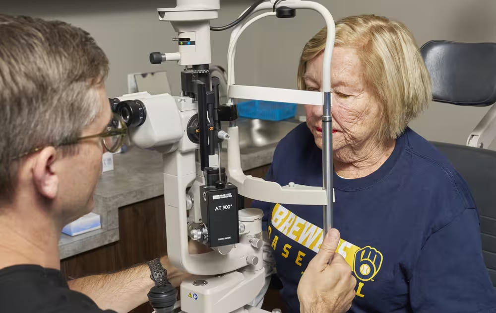
Macular degeneration, also known as age-related macular degeneration (AMD), is one of the leading causes of vision loss among adults over the age of 50. It affects the macula, the central part of the retina responsible for sharp, detailed vision needed for activities such as reading, driving, and recognizing faces. AMD can severely impact your quality of life if it progresses and/or is left untreated. Understanding the two main forms – dry and wet macular degeneration – is key to early detection and preserving your vision. What Is Dry Macular Degeneration? Dry macular degeneration is the more common form, accounting for about 85-90% of AMD cases. It develops gradually as the light-sensitive cells in the macula and supporting tissues break down over time. Key Features: Slow, progressive blurring of central vision Presence of drusen, small yellow deposits under the retina Gradual thinning of the macula Usually affects both eyes, though one may progress faster than the other Symptoms: Blurry or distorted central vision Colors appearing less bright or vivid Need for brighter light when reading or performing close work Dry AMD can progress throughout your life and, if severe, can lead to atrophy (scarring) of the macula. This can significantly affect your central vision and is irreversible. What Is Wet Macular Degeneration? Wet macular degeneration is less common but more serious. It occurs when abnormal blood vessels grow beneath the retina, leaking fluid or blood and causing rapid damage to the macula. Key Features: Sudden and severe vision loss Distortion of straight lines (appearing wavy or bent) Rapid central vision changes If you notice these symptoms, it’s essential to seek immediate ophthalmic care – early intervention can make a significant difference in improving or preserving vision. Who is at Risk for Macular Degeneration? Several factors can increase the likelihood of developing AMD: Age: Most common in people over 50 Genetics: Family history of AMD increases risk Smoking: You are 20x more likely to develop AMD if you smoke Sun Exposure: Prolonged UV exposure may contribute to retinal damage How is Macular Degeneration Treated? Macular degeneration is a lifelong, progressive condition. Currently, there is no cure for this disease, but there are interventions that can slow disease progression. Treating Dry Macular Degeneration: Nutritional Supplements: The AREDS2 (Age-Related Eye Disease Study 2) formula – containing vitamins C and E, zinc, copper, lutein, and zeaxanthin – can help reduce the risk of AMD progressing. Healthy Diet: Focus on leafy greens, colorful fruits and vegetables, and fish rich in omega-3s. Lifestyle Changes: Stop or cut back on smoking and protect eyes from UV light with sunglasses outside. Regular Monitoring: Frequent eye exams allow for early detection of any transition to the wet form. Complement Inhibitor Injections: For advanced, severe, dry AMD with macular atrophy (scarring), injections of medications such as pegcetacoplan (Syfovre) and avacincaptad pegolin (Izervay) may help slow down the progression of atrophy. Treating Wet Macular Degeneration: In addition to the above listed treatments for dry AMD, wet AMD can be treated with: Anti-VEGF Injections: Medications such as bevacizumab (Avastin), aflibercept (Eylea), or faricimab (Vabysmo) block abnormal blood vessel growth and leakage. Wait – Injections into my Eye? Yes! While it can sound intimidating to receive an eye injection, these medications work very well to improve or maintain your vision. For an injection, your eye is numbed with eyedrops and then with local anesthetic. Antiseptic eyedrops are used to ensure that the eye surface is clean and minimize infection risk. Most patients do not feel the injection or only feel slight pressure with the injection. After the injection, the eye may feel slightly scratchy for the rest of the day and you may see a new floater in your vision, but this improves quickly. How Can I Protect my Vision? Regular comprehensive eye exams are the best defense against vision loss from macular degeneration. At Ophthalmology Associates, S.C., our ophthalmologists use advanced diagnostic imaging and personalized care plans to detect AMD early and manage it effectively. If you’ve noticed changes in your central vision or have risk factors for macular degeneration, schedule an eye exam today. Early diagnosis and treatment can help preserve your sight for years to come. To check your vision at home, you can use the Amsler Grid once or twice a week. Wear your reading glasses if you normally use them. Hold the grid about 12–14 inches (30–35 cm) from your face in good lighting. Cover one eye and focus on the center dot with the uncovered eye. While keeping your gaze fixed on the dot, notice whether: Any lines appear wavy, blurred, or distorted Any areas of the grid look dark, blank, or missing Repeat with the other eye. If you notice any new or worsening distortions, blurring, or missing areas, contact your ophthalmologist immediately. These changes may indicate the development or progression of wet macular degeneration, which requires prompt treatment to preserve vision. Print Your Own Amsler Grid: https://www.amslergrid.org/AmslerGrid.pdf Book an Appointment
Ophthalmology Associates, S.C. Wins Milwaukee’s Top Choice Awards 2025

We are proud to announce that Ophthalmology Associates, S.C. has been honored with the Milwaukee Top Choice Awards 2025 for the second time in a row! This recognition is a true reflection of the trust our community places in us and the dedication our team brings to eye care every single day. A Recognition of Excellence in Eye Care Winning this award underscores our dedication to delivering the highest quality ophthalmology services in Milwaukee and its surrounding areas, including Greenfield, Cudahy, and New Berlin. From comprehensive eye exams to advanced treatments for cataracts, glaucoma, and retinal conditions, our board-certified ophthalmologists and optometrists work tirelessly to ensure every patient receives personalized, compassionate care. What the Award Means to Us The Milwaukee Top Choice Awards are a community-driven recognition, where patients and residents vote for the businesses they value most. Being chosen as the top eye care provider in Milwaukee means more than just an award – it’s a reminder that the work we do impacts lives every day. Our patients trust us with one of their most valuable senses – vision – and this award reaffirms our responsibility to continue raising the standard of care. Thank You to Our Patients and Team We extend heartfelt gratitude to our patients for their loyalty and confidence in our services. Your trust and support inspire us to continuously invest in the latest technology and treatments to better serve you. A special thank you also goes to our incredible team of doctors, optometrists, technicians, and support staff, whose expertise and dedication make recognitions like this possible. Looking Ahead As we celebrate this achievement, our mission remains the same: to help our patients see the world more clearly and live life to the fullest. We are excited about the future and look forward to continuing to serve Milwaukee with excellence in eye care for many years to come. Schedule your next appointment with Ophthalmology Associates, S.C. today and experience award-winning eye care trusted by the Milwaukee community. Book an Appointment